Eye Examination Simulator
Product Supervision:
Japan society for Medical Education Working Group with the cooperation of: Kansai Medical University Department of Ophthalmology
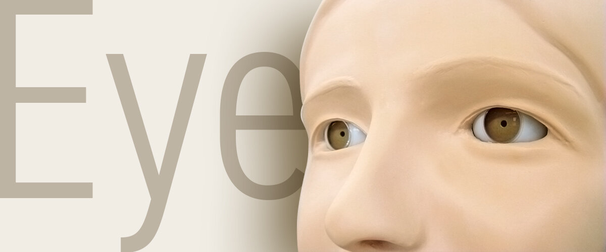
Longtime Bestseller!
Practical fundus examination simulator with 10 clinical images and variations
TRAINING SKILLS
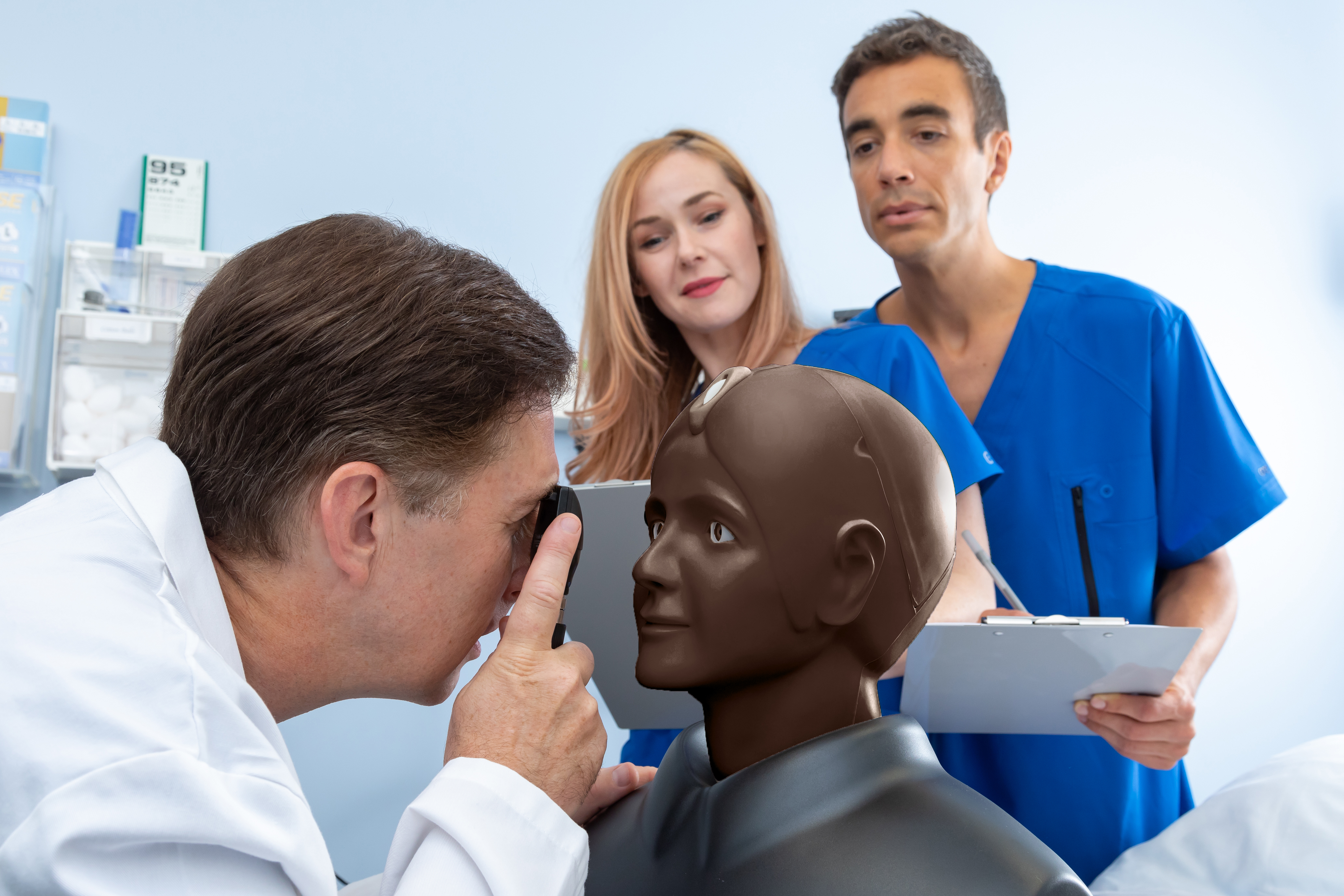
- Fundus examination with direct ophthalmoscope
- Differentiation pathologies
- Communication
- Correct use of direct Ophtalmoscope
FEATURES
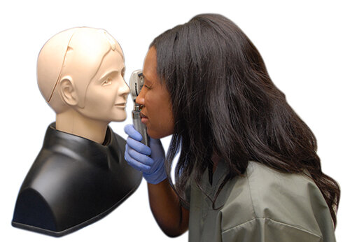 Lens-Equipped eyeball units which simulates visual axis of human eye
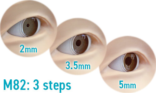 Different pupil sizes can be set - 3 steps (2, 3.5, 5mm dia.) in M82, 2 steps(3.5, 8mm dia.) in M82A for challenge variation
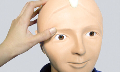 Realistic raising of Eyelid
Practice how to position your subdominant hand and your body against the patient.
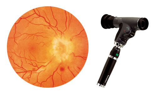 Detectable Red Reflex
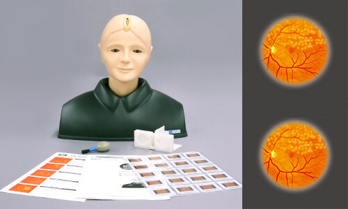 Most common pathologies with in 10 real clinical images
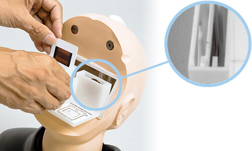 Adjustable fundus depth : Normal / Myopia / Hyperopia
Highly recommended for
-Undergraduate Medical Students
-OSCE
-Training for Nurse Practitioners
-Training for General Practitioners
CASES

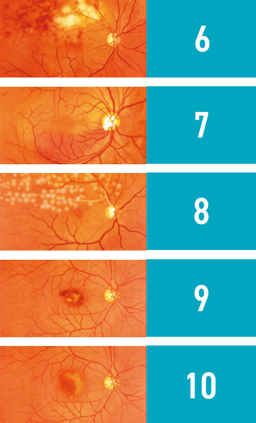
1 | Normal fundus
2 | Hypertensive retinopathy:
- Grade 3 arteriolar vasoconstriction
- Grade 1 arteriolosclerosis
- hemorrhages and cotton wool spots
- simple vein concealment
3 | Diabetic retinopathy:
microaneurysm, hemorrhages and hard exudates
4 | Papilloedema (chronic phase)
5 | Papilloedema (acute phase)
6 | Glaucomatous optic atrophy:
glaucomatous optic disc cupping and nerve fiber defect
7 | Retinal vein occlusion (acute phase):
flame-shaped hemorrhage and cotton wool spots
8 | Retinal vein occlusion (post retinal laser photocoagulation)
9 | Toxoplasmosis: retinochoroiditis
10| Age-related macular degeneration:
macular exudates and subretinal hemorrhage
*M82A includes two normal fundus slides.

Difference of M82 (3 steps) and M82A (2 steps)
Pupils of M82A are designed larger than M82.
M82A: 3.5, 8mm dia.
M82: 2, 3.5, 5mm dia.
This offers advantages for providing;
-dilated pupils when using mydriaticeye drops
-visual intelligibility for bigginers